CASE 163
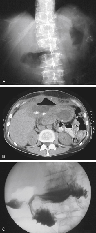
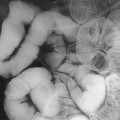
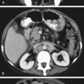
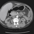
History: A 55-year-old man presents with abdominal pain.
1. What should be included in the differential diagnosis of the imaging finding shown in Figures A and B? (Choose all that apply.)
2. Which of the following is a mucosal defect replacing two thirds of the duodenal bulb?
3. Where do perforating ulcers most commonly occur?
Stay updated, free articles. Join our Telegram channel

Full access? Get Clinical Tree


