CASE 164
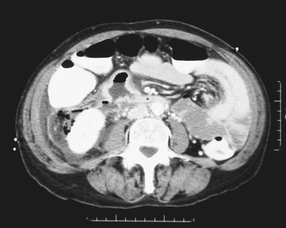
History: A 51-year-old woman presents with right-sided abdominal pain, nausea, and vomiting of brackish dark emesis. She is a long-term user of aspirin and Goody’s Powder pain reliever.
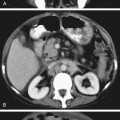
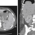
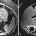
1. What should be included in the differential diagnosis of the imaging finding shown in the figure? (Choose all that apply.)
D. Emphysematous pyelonephritis
E. Post–endoscopic retrograde cholangiopancreatography (ERCP) and postsphincterotomy
2. Which statement regarding possible causes of duodenal perforation is true?
A. Duodenal diverticula are most commonly bulbar.
B. Blunt force trauma usually does not cause duodenal perforation.
C. Duodenal perforation after ERCP and sphincterotomy is mostly managed surgically.
D. Peptic ulceration is diagnosed at CT by the presence of circumferential wall thickening.
Stay updated, free articles. Join our Telegram channel

Full access? Get Clinical Tree


