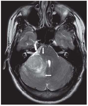
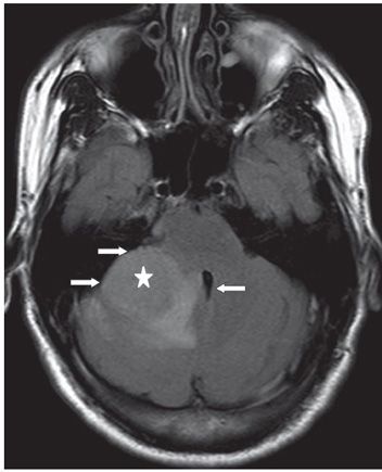
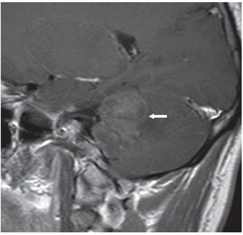
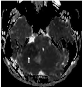
FINDINGS Figure 164-1. Axial NCCT through the posterior fossa. There is a large hyperdense mass (vertical arrow) laterally on the right in the posterior fossa. There is hypodensity medially to the mass (transverse arrow) consistent with edema. Figure 164-2. Axial T2WI through the mass. The mass is mildly heterogeneously hyperintense and exophytic with widening of the right cerebellopontine angle (CPA) and normal-appearing right internal auditory canal (IAC) (chevron). There is compression of the right brachium pontis (vertical arrow). There is edema medially (transverse arrow). Figure 164-3. Axial FLAIR through the mass. The round mass is mildly hyperintense (star) with surrounding edema medially and compression of the fourth ventricle (single arrow) and brainstem. There is scalloping of the right petrosal surface (lateral 2 arrows over the petrous bone). Figure 164-4. Sagittal post-contrast T1WI through the mass. There is diffuse contrast enhancement of the mass (arrow). Figures 164-5 and 164-6


Stay updated, free articles. Join our Telegram channel

Full access? Get Clinical Tree


