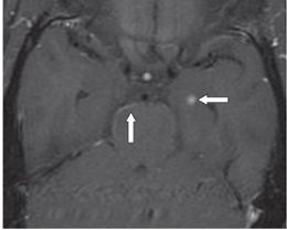
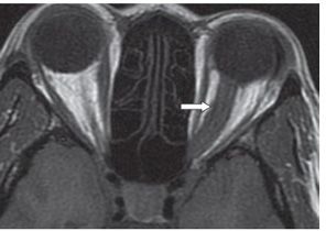
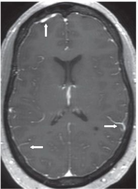
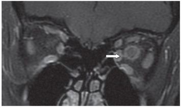
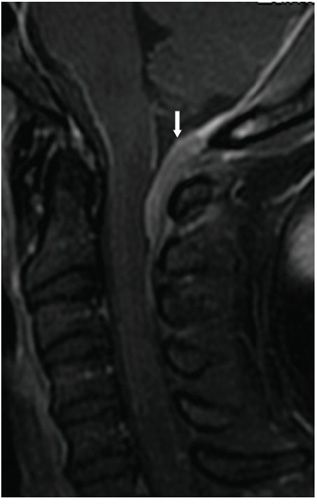
FINDINGS Figures 165-1. Axial MR T2WI through the corona radiata. There are multifocal small hyperintense lesions mainly in the deep white matter of bilateral cerebral hemispheres (arrows). Figure 165-2. Axial post-contrast T1WI through the middle cranial fossae. There is a small left medial temporal focal contrast enhancement (transverse arrow). There is mild leptomeningeal enhancement around the brainstem (vertical arrow). Figure 165-3. Axial non- contrast T1WI through the orbits. There is thickened hypointense left optic nerve and sheath (arrow). Figure 165-4. Axial post-contrast T1WI through the lateral ventricles. There is multifocal leptomeningeal and pachymeningeal enhancement (arrows). Figure 165-5. Coronal post-contrast T1WI with fat suppression of the orbits. The left optic nerve and sheath show enlargement and enhancement (transverse arrow). Figures 165-6. Sagittal post-contrast T1WI, through the foramen magnum. There is thickened contrast-enhancing dural plague (pachymeninges) posteriorly at the craniovertebral junction (arrow) projecting into the cerebrospinal fluid (CSF) space.
DIFFERENTIAL DIAGNOSIS


Stay updated, free articles. Join our Telegram channel

Full access? Get Clinical Tree


