CASE 166
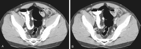
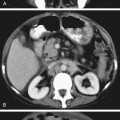
History: A 39-year-old man presents with left lower quadrant pain of 4 days’ duration.
1. What should be included in the differential diagnosis of the imaging finding shown in Figure A? (Choose all that apply.)
2. What is the etiology of epiploic appendagitis?
A. Occlusion of the lumen of the appendage with debris
B. Bacterial contamination of the epiploic appendage
C. Torsion of the epiploic appendage
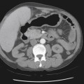
3. The lesion of epiploic appendagitis is described as an oval pericolonic lesion surrounded by inflammatory stranding. What is the density of this oval lesion?
Stay updated, free articles. Join our Telegram channel

Full access? Get Clinical Tree


