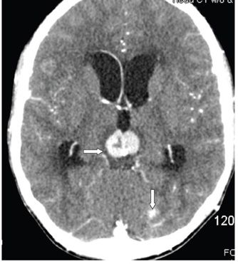
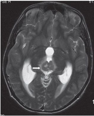
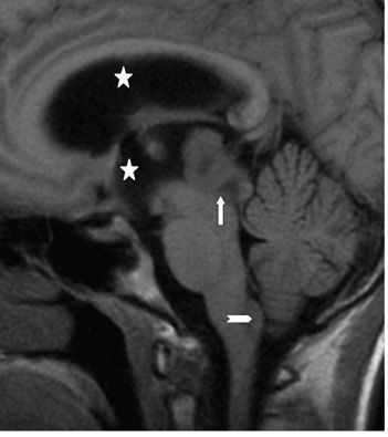

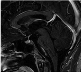
FINDINGS Figures 166-1 and 166-2. Axial pre- and post-contrast CT, respectively, through the pineal region. There is a 2-cm well-circumscribed contrast-enhancing hyperdense mass in the pineal region (arrow). A tiny calcification is present posteriorly in the mass. There are three other similar lesions in the suprasellar cistern (not shown), along the left tentorium (vertical arrow) and in the inferior fourth ventricle (not shown). There is hydrocephalus (stars). Figure 166-3. Axial T2WI through the pineal region. There is a well-circumscribed isointense mass (to gray matter [GM]) with three tiny hyperintense foci internally in the pineal region (arrow). There is surrounding hyperintensity in the thalami suggesting edema. There is hydrocephalus. Figures 166-4 and 166-5. Pre- and post-contrast sagittal T1WI through the pineal mass. The masses are isointense to hypointense with avid contrast enhancement in the pineal region (vertical arrow), hypothalamus (transverse arrow), and at the obex (chevron). The stars are within dilated third and lateral ventricles consistent with hydrocephalus. Figure 166-6


Stay updated, free articles. Join our Telegram channel

Full access? Get Clinical Tree


