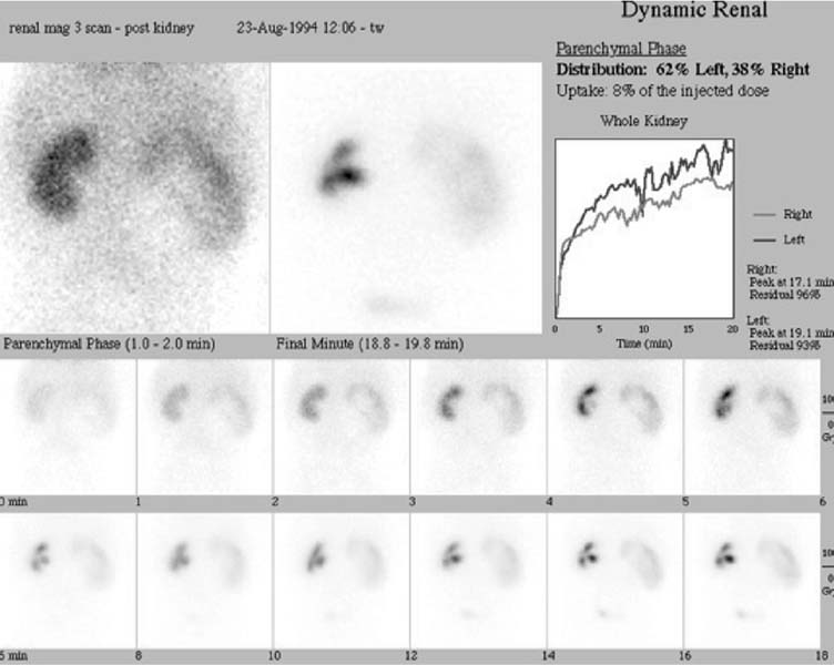
CASE 166 A 7-week-old infant boy presents with severe right hydronephrosis detected by prenatal ultrasonography. Postnatal ultrasonography also demonstrates severe pelvicaliectasis of the right collecting system, consistent with right ureteropelvic junction obstruction. Voiding cystourethrography shows no reflux. A 99mTc-MAG-3 study is obtained to evaluate for the presence and severity of obstruction. Fig. 166.1 Fig. 166.2 • 99mTc-MAG-3 is administered at a dose of 0.2 mCi/kg. The minimum dose is 2 mCi; the maximum dose is 10 mCi. • Use a high- or ultra-high-resolution, low-energy, parallel-hole collimator. • Energy window is 20% centered at 140 keV. • Dynamic images are acquired for a minimum of 20 minutes (or until tracer is seen in the collecting system). For the diuretic phase, after the administration of furosemide (dose, 1 mg/kg; maximum dose, 40 mg), dynamic images are acquired for 30 minutes. • Bladder catheterization and intravenous access are established before the injection of radiotracer. The parenchymal phase (Fig. 166.1) shows an enlarged right kidney with a central area of decreased tracer uptake representing the dilated collecting system. The relative uptake of radiotracer is 38% by the right kidney and 62% by the left kidney. The cortical transit time, defined as the time between the injection of tracer and the first appearance of tracer in the collecting system, is longer than 20 minutes for the right side and 4 to 5 minutes for the left. There is marked retention of tracer in the right collecting system before administration of the diuretic. Thirty minutes after the administration of furosemide (Fig. 166.2), there is 91% residual radiotracer in the right pelvis, and a half-time of longer than 100 minutes is noted. These findings indicate a right ureteropelvic junction obstruction. Tracer washout from the left kidney is augmented by furosemide, indicating no obstruction. • Obstructed urinary collecting system
Clinical Presentation
Technique
Image Interpretation
Differential Diagnosis
Stay updated, free articles. Join our Telegram channel

Full access? Get Clinical Tree


