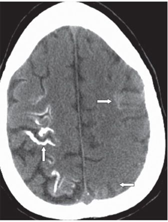
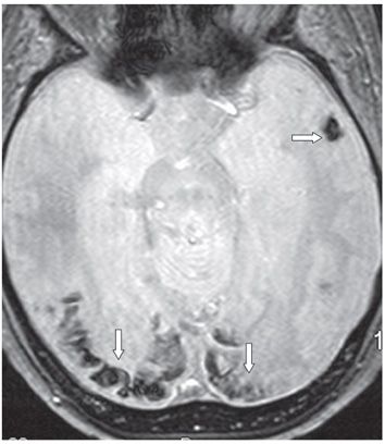
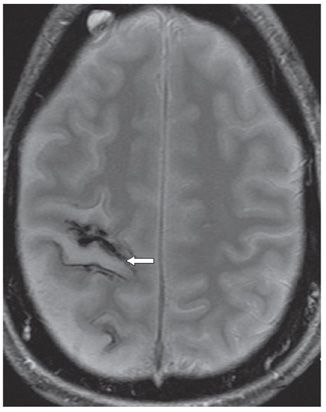
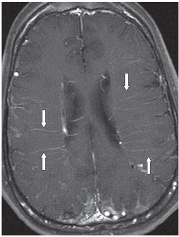
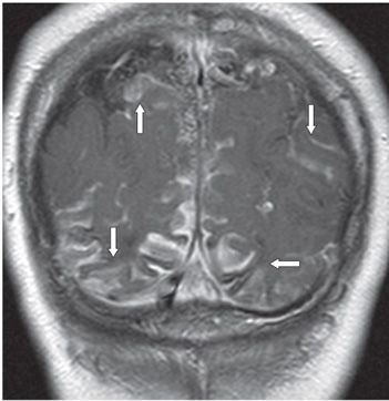
FINDINGS Figure 167-1. Axial NCCT through the occipital lobes. There are bilateral occipital cortical calcifications (arrows) extending to the posterior right temporal lobe. Figure 167-2. Axial NCCT through the frontoparietal convexity. There are right frontotemporal gyriform (tramline) calcifications (vertical arrow). Some faint left frontoparietal cortical calcifications are present (transverse arrows). Figure 167-3. Axial GRE through the occipital lobes. There is bilateral occipital cortical hypointensity/blooming in the areas of calcifications seen in Figure 167-1. There is a small focus of left temporal lobe hypointensity (arrow). Figure 167-4. Axial GRE through the frontoparietal convexities. Right frontoparietal multifocal cortical hypointensity/blooming is present. There is dilatation of the right central sulcus (arrow) consistent with volume loss. Figure 167-5. Axial post-contrast T1WI through the corona radiata. There are prominent transmedullary veins coursing through the corona radiata bilaterally (arrows). Figure 167-6


Stay updated, free articles. Join our Telegram channel

Full access? Get Clinical Tree


