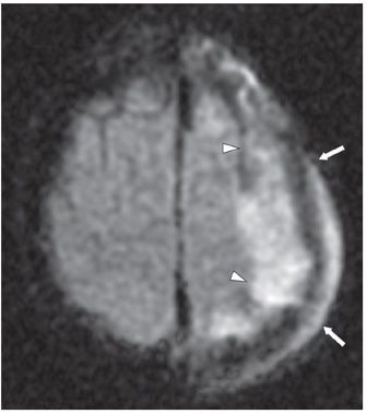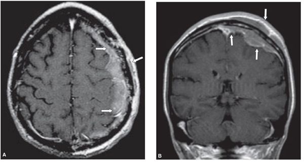

FINDINGS Figure 168-1. Sagittal T2WI. There is abnormal, lobular isointense extraaxial tissue along the inner table of the skull (arrowheads). There is infiltration of the bony calvarium with contiguous involvement of the deep scalp layers (arrows). Figure 168-2. Axial trace DWI. There is restricted diffusion within the intracranial (arrowheads) and scalp components (arrows) of the mass (confirmed on ADC, not shown). Figure 168-3. Axial post-contrast T1WI. There is diffuse enhancement of the extraaxial and extracranial components of the mass along with permeative destruction of the intervening frontoparietal calvarium (arrows).
DIFFERENTIAL DIAGNOSIS Subdural empyema/abscess with calvarial osteomyelitis and scalp infection, dural/calvarial lymphoma, dural/calvarial metastasis, aggressive meningioma, plasmacytoma.
DIAGNOSIS Secondary intracranial lymphoma.
DISCUSSION
Stay updated, free articles. Join our Telegram channel

Full access? Get Clinical Tree


