Case 169
Case History
A 55-year-old woman presents with a left breast lump. She has had bilateral augmentation mammoplasty. This is her baseline examination.
Physical Examination
• left breast: mass on medial breast
• right breast: normal exam
Mammogram
Mass
• margin: spiculated
• shape: irregular
• density: equal density
Calcifications (Fig. 169–1)
• type: pleomorphic/heterogeneous
• distribution: grouped/clustered
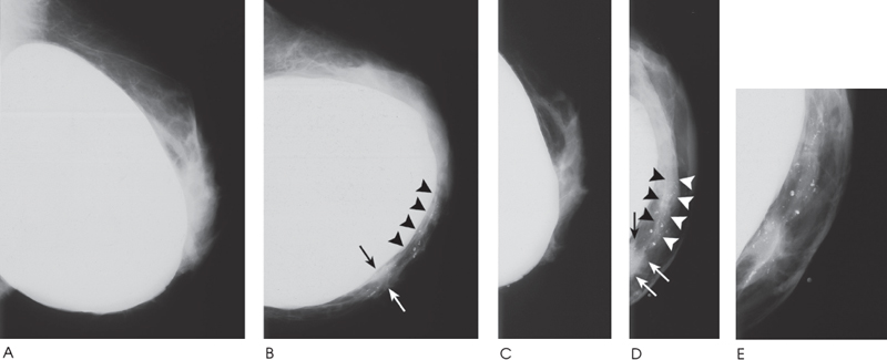
Figure 169–1. In the left medial breast there is an irregular density (arrows) containing heterogeneous calcifications. Adjacent to the mass are more heterogeneous calcifications (arrowheads). (A). Left MLO implant mammogram. (B). Left CC implant mammogram. (C). Left MLO implant displaced mammogram. (D). Left CC implant displaced mammogram. (E). Close-up of left medial breast.
Ultrasound
Low Frequency
Frequency
• 7.5 MHz
Mass
• margin: ill defined
• echogenicity: hypoechoic
• retrotumoral acoustic appearance: no shadowing
• shape: irregular (Fig. 169–2)


Stay updated, free articles. Join our Telegram channel

Full access? Get Clinical Tree


