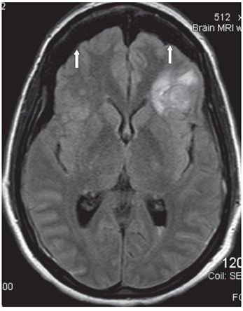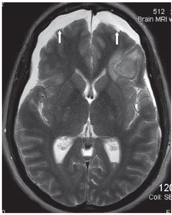

FINDINGS Figure 169-1. NCCT through the inferior frontal lobes. There is extraaxial crescentic large cerebrospinal fluid (CSF) density space measuring about 1 cm thick over both frontal lobes with effacement of underlying sulci (vertical arrows). There is associated left frontal opercula hematoma (transverse arrow) and IVH in left trigone (posterior left arrow). Figures 169-2 and 169-3. Axial FLAIR and T2WI through the frontal lobes respectively. The bifrontal extraaxial collections have CSF intensity on all sequences (arrows). The left frontal opercula hematoma and IVH are again visualized.
DIFFERENTIAL DIAGNOSIS Atrophy or volume loss, subdural hygroma (SDG), chronic subdural hematoma (CSDH).
DIAGNOSIS Traumatic subdural hygroma (TSDG).
DISCUSSION
Stay updated, free articles. Join our Telegram channel

Full access? Get Clinical Tree


