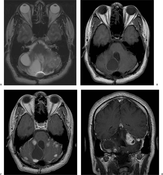
Case 17 A 35-year-old man with the history of a resected tumor in the cerebellum presenting with worsening dysmetria. (A) Axial T2-weighted image (WI) demonstrates multiple cystic lesions (asterisks) in the cerebellum bilaterally and areas of encephalomalacia on the left side. Note the magnetic susceptibility artifact near the occipital bone resulting from prior surgery (arrow). (B) Axial T1WI of the brain without contrast showing well-defined cystic lesions with signal slightly higher than that of cerebrospinal fluid (arrows). (C) Axial postcontrast T1WI shows numerous enhancing solid lesions (arrow) and a mural nodule (asterisk) along the wall of one of the cerebellar cysts. (D) Coronal postcontrast T1WI shows numerous enhancing solid lesions (arrows
Clinical Presentation
Imaging Findings
![]()
Stay updated, free articles. Join our Telegram channel

Full access? Get Clinical Tree


