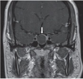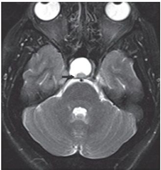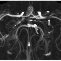

FINDINGS Figure 17-1. Sagittal T1WI. There is an intrasellar mass that is isointense with the brain (arrow). The hyperintense neurohypophysis is not present. Figure 17-2. Coronal post-contrast T1WI through the sella turcica. The mass does not enhance but its periphery does (arrow) probably due to surrounding normal pituitary tissue. Figure 17-3. Axial T2WI through the sella. There is a posterior mural nodule (arrow) that is typical of this entity and helps differentiate it from other cystic pituitary masses.
DIFFERENTIAL DIAGNOSIS Rathke cleft cyst, cystic pituitary adenoma, craniopharyngioma.
DIAGNOSIS Rathke cleft cyst.
Stay updated, free articles. Join our Telegram channel

Full access? Get Clinical Tree


