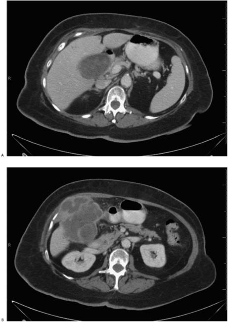
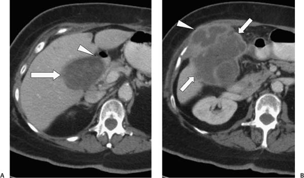
Case 17 A 69-year-old woman presents with right upper quadrant pain. (A) Contrast-enhanced computed tomography shows a multilobular soft-tissue mass occupying most of the lumen of the gallbladder (GB). The GB wall is thickened (arrow) and compressing the adjacent duodenum (arrowhead). (B) A more caudal image shows a multiloculate cystic mass (arrows) surrounding the thickened GB, which is seen posteromedially. This mass may be invading the abdominal wall musculature (arrowhead). • GB adenocarcinoma: A noncalcified, multilobular mass effacing the GB lumen, GB wall thickening, and an exophytic mass involving the GB and invading the abdominal wall are all features of GB carcinoma. • Xanthogranulomatous cholecystitis (XGC): A multiloculated mass involving the GB and invading the abdominal wall is suspicious for this entity. A more characteristic appearance would include hypodense nodules in the GB wall and stones. • Metastatic disease:
 Clinical Presentation
 Imaging Findings
 Differential Diagnosis
![]()
Stay updated, free articles. Join our Telegram channel

Full access? Get Clinical Tree


