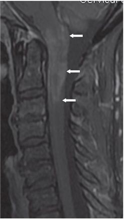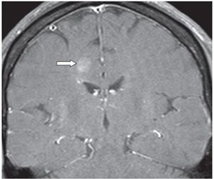

FINDINGS Figure 170-1. Sagittal short tau inversion recovery (STIR) cervical spine MRI. There is enlargement of the cervical spinal cord with smudgy hyperintensity from the level of the foramen magnum to the C4 level (arrows). Hyperintensity extends to the medulla. Figure 170-2. Sagittal post-contrast T1WI through the cervical spine. There is smudgy contrast enhancement within the lesion from the level of the foramen magnum to C3 (arrows), short of the inferior extent of the lesion on STIR. Lesion is isointense without contrast (not shown). Figure 170-3. Coronal post-contrast T1WI through the level of the third ventricle. There is a poorly marginated round contrast-enhancing lesion in parasagittal right posterior frontal centrum semiovale (arrow). There were a few other white matter lesions elsewhere (not shown).
DIFFERENTIAL DIAGNOSIS Longitudinally extensive transverse myelitis (LETM), neuromyelitis optica (NMO), multiple sclerosis (MS), astrocytoma, ependymoma.
DIAGNOSIS Neuromyelitis optica (NMO).
DISCUSSION
Stay updated, free articles. Join our Telegram channel

Full access? Get Clinical Tree


