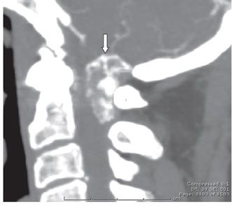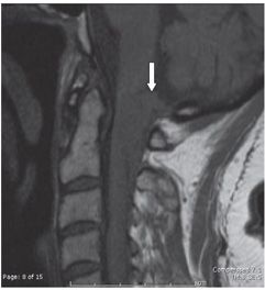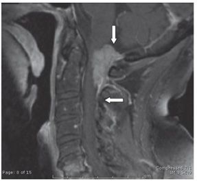


FINDINGS Figures 172-1 and 172-2. Axial NCCT brain and sagittal MIP reformatted images, respectively, from a head and neck CTA. There is a partially calcified (peripherally) extraaxial dural-based lesion arising from the posterior aspect of the foramen magnum (arrows) compressing the upper spinal cord. Figures 172-3 and 172-4. Sagittal pre- and post-contrast T1WI, respectively. There is a homogeneously contrast-enhancing (Figure 172-4) isointense (Figure 172-1) dural-based lesion at the posterior aspect of the foramen magnum (vertical arrows) extending inferiorly to C2 level with anterior displacement and compression of the adjacent medulla and upper cervical cord. There is a short dural tail inferiorly in Figure 172-4 (transverse arrow).
DIFFERENTIAL DIAGNOSIS Meningioma, dural metastasis, granulomatous processes such as sarcoidosis and tuberculosis (TB), primary bone neoplasm, that is, osteogenic sarcoma, chondrosarcoma.
DIAGNOSIS Foramen magnum meningioma.
DISCUSSION
Stay updated, free articles. Join our Telegram channel

Full access? Get Clinical Tree


