Courtesy of Annette Douglas, MD.
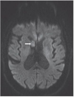
Courtesy of Annette Douglas, MD.
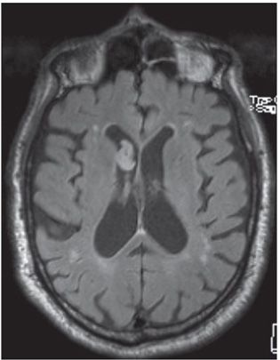
Courtesy of Annette Douglas, MD.
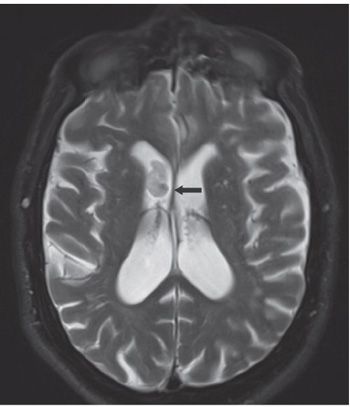
Courtesy of Annette Douglas, MD.
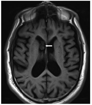
Courtesy of Annette Douglas, MD.
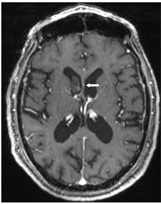
Courtesy of Annette Douglas, MD.
FINDINGS Figures 173-1 and 173-2. Axial ADC map and corresponding DWI through the lateral ventricles. There is a smooth nodular mass within the right frontal horn showing no evidence of restricted diffusion (arrows). Figures 173-3 and 173-4. Axial FLAIR and T2WI, respectively, through the lateral ventricles. There is a smooth marginated mildly hyperintense right ventricular nodular mass with a tiny focal hypointensity posteromedially on the T2WI (arrow) (focal calcification or hemorrhage?). There is no evidence of perilesional edema or mural attachment. Figures 173-5
Stay updated, free articles. Join our Telegram channel

Full access? Get Clinical Tree


