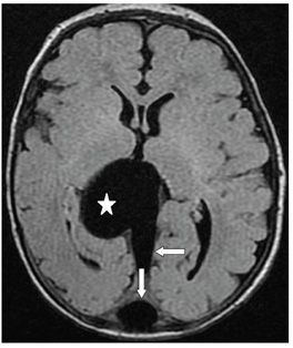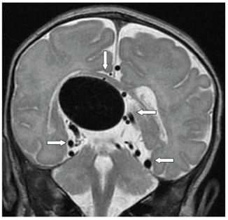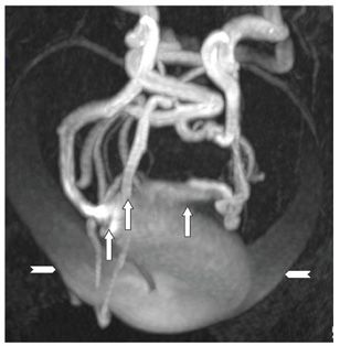


FINDINGS Figure 174-1. Sagittal Doppler cranial ultrasound. There is a large 3.5-cm vascular structure posteriorly to the third ventricle and splenium of corpus callosum (appropriate position of the vein of Galen) (star) with two large feeding vessels demonstrated: a pericallosal artery and a posterior cerebral artery (arrows). Figure 174-2. Axial FLAIR through the third ventricle. There is a 3.2 cm × 3.1 cm signal void posteriorly to the thalami and third ventricle with mass effect consistent with the vein of Galen aneurysmal malformation (VGAM) (star). Large draining sinus (transverse arrow) and torcula/SSS (vertical arrow) are seen posteriorly to the aneurysm. Figure 174-3. Coronal T2WI through the aneurysm. There is superior displacement of the splenium of the corpus callosum (vertical arrow). Multiple round and tubular signal voids representing large afferent arteries (arrows) are seen surrounding the aneurysm. Figure 174-4. 3D TOF MRA of the head Submentovertical (SMV) view. There is direct connection of all major intracranial vessels (arrows), anterior cerebral artery (ACA), posterior cerebral artery (PCA), and middle cerebral artery (MCA) to the aneurysmal malformation. Huge bilateral transverse sinuses (chevrons) emanate from the torcula.
DIFFERENTIAL DIAGNOSIS N/A.
DIAGNOSIS VGAM.
Stay updated, free articles. Join our Telegram channel

Full access? Get Clinical Tree


