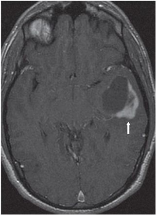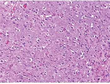

FINDINGS Figure 175-1. Axial T2WI through the temporal lobes. There is a 4-cm relatively sharply circumscribed mass in the left temporal lobe. The anterior cystic component has cerebrospinal fluid (CSF) hyperintensity with the solid posterior component showing a slightly less hyperintense signal character (vertical arrow). There is minimal surrounding smudgy hyperintensity posteromedially (transverse arrow) that is consistent with edema. There is moderate mass effect with effacement of the overlying gyri and a moderate amount of medial shift of the uncus. Figure 175-2. Axial post-contrast T1WI. This shows the anterior cystic component with enhancing thick posterolateral solid component (arrow). Figure 175-3. Photomicrograph shows tumor composed of haphazardly arranged ganglion/ganglioid cells in a neuropil-rich stroma. Few eosinophilic granular bodies (EGBs) are also noted. Glial cells are not prominent in this area (H&E stain).
DIFFERENTIAL DIAGNOSIS Ganglioglioma (GG), dysembryoplastic neuroepithelial tumor (DNET), pleomorphic xanthoastrocytoma (PXA), metastatic lesion, tumefactive demyelination, oligodendroglioma, astrocytoma.
DIAGNOSIS Ganglioglioma (GG).
DISCUSSION
Stay updated, free articles. Join our Telegram channel

Full access? Get Clinical Tree


