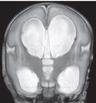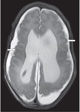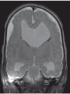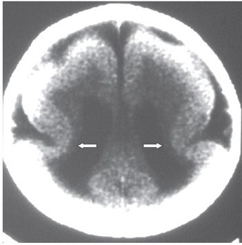


FINDINGS Figure 176-1. Axial T2WI through the lateral ventricles. There is dilatation of the lateral ventricles (stars) consistent with hydrocephalus. There is smooth brain surface (no sulci visible) with shallow sylvian fissures (arrows) and hourglass configuration of the cerebral hemispheres. Figure 176-2. Coronal T2WI through the sylvian fissures. Hydrocephalus with shallow sylvian fissures and smooth brain surface again visualized. Figures 176-3 and 176-4. Axial and coronal T2WI through the lateral ventricles following shunting of the hydrocephalus. Convexity sulci have appeared (arrows); rather shallow with small gyri. There is hypoplasia of the periventricular white matter (WM). Figure 176-5
Stay updated, free articles. Join our Telegram channel

Full access? Get Clinical Tree


