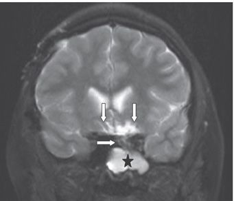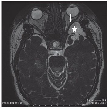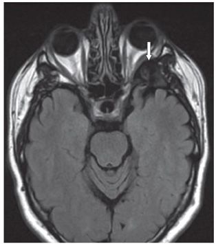


FINDINGS Figure 177-1. Axial CTA image, bone window through the ethmoid. There is comminuted ethmoidal fracture. Additionally, there is a fracture through the sphenoid roof at the skull base with pneumocephalus in the region of the suprasellar cistern (arrows). Figure 177-2. Coronal T2WI through the planum sphenoidale 1 year later. There is encephalomalacia and gliosis of the bilateral subfrontal lobes (vertical arrows). Additionally, there is hyperintense opacification in the region of the posterior ethmoid and sphenoid sinuses (star). There appears to be a tenuous tract through the planum on the left (transverse arrow). Figures 177-3 and 177-4. Axial heavily T2WI fast imaging employing steady state acquisition (FIESTA) and FLAIR, respectively, in a companion case. There is hyperintense collection anterior to the left middle cranial fossa (star) through a defect in the dura and the left greater wing of sphenoid. Fluid collection abuts and compresses the left lateral rectus (arrows). The fluid suppresses on FLAIR in Figure 177-4.
DIFFERENTIAL DIAGNOSIS Growing skull fractures, pseudomeningocele, leptomeningeal cyst, sinus retention cyst and infection.
DIAGNOSIS Posttraumatic cerebrospinal fluid (CSF) collection (pseudomeningocele).
DISCUSSION
Stay updated, free articles. Join our Telegram channel

Full access? Get Clinical Tree


