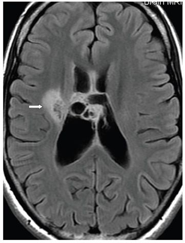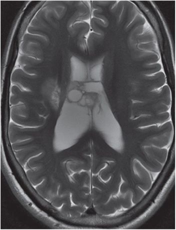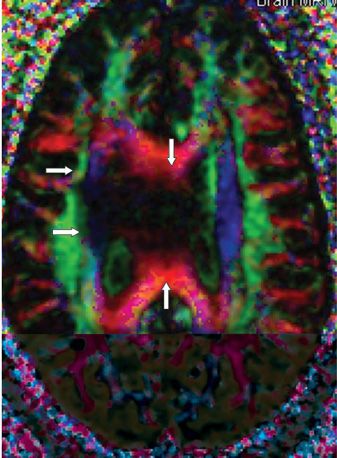


FINDINGS Figure 179-1. Sagittal T1WI through the corpus callosum (CC). There is a mixed cystic anteriorly (cerebrospinal fluid [CSF] intensity) and solid hypointense (posteriorly) mass in the midbody of the CC (arrow) projecting to the septum pellucidum inferiorly. Post-contrast T1WI (not shown) demonstrated very minimal contrast enhancement. Figure 179-2. Axial FLAIR through the corona radiata. The solid components are hyperintense and project into the right corona radiata (arrow), while the cystic components show CSF hypointensity. Figure 179-3. Axial T2WI through the corona radiata. The solid components are somewhat hyperintense but brighter than surrounding white matter (WM). Figure 179-4. Axial DTI color directional map through the CC. There is disruption of the red CC fibers (vertical arrows) and the adjacent right superior fronto-occipital fasciculus and the superior internal capsule (transverse arrows).
DIFFERENTIAL DIAGNOSIS Glioblastoma (GB), lymphoma, tumefactive or toxic demyelination, oligodendroglioma.
DIAGNOSIS CC oligodendroglioma II (ODG II) recurrent.
DISCUSSION
Stay updated, free articles. Join our Telegram channel

Full access? Get Clinical Tree


