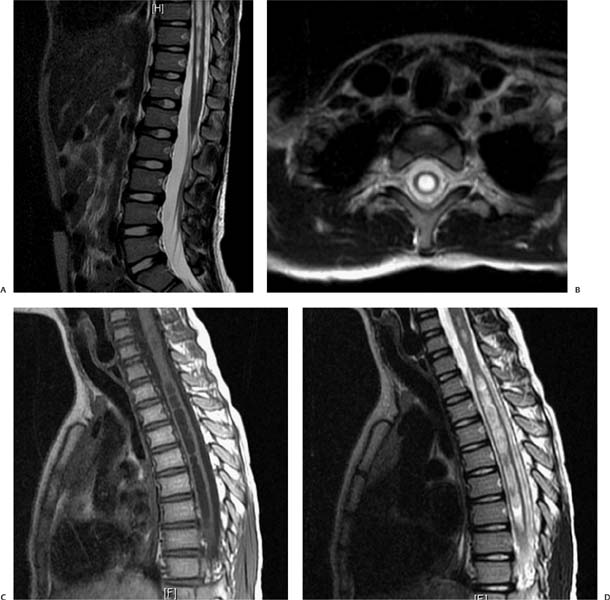
Case 18 A 7-year-old boy with neck paresthesias and hand tingling. (A) Sagittal T2-weighted image (WI) of the lower spine demonstrates a well-defined central area of fluid signal within the cord, which tapers toward the conus medullaris (arrow). (B) Axial T2WI in the cervical region shows a central fluid cavity within the cord with well-defined margins (arrow). There is mild cord expansion. (C) Sagittal T1WI shows extension of the cystic cavity to the cervical cord and multiple septa (arrowheads). (D) Sagittal T2WI of the upper spine demonstrates expansion of the cord in the area that contains the fluid cavity, with a waist in the region where the cord is normal (arrowhead). • Syringohydromyelia: This is characterized by a longitudinally oriented cavity within the spinal cord, with cerebro-spinal fluid (CSF) signal on all the sequences. There is no solid component or enhancement.
Clinical Presentation
Imaging Findings
Differential Diagnosis
Stay updated, free articles. Join our Telegram channel

Full access? Get Clinical Tree


