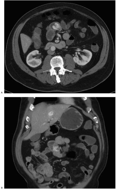
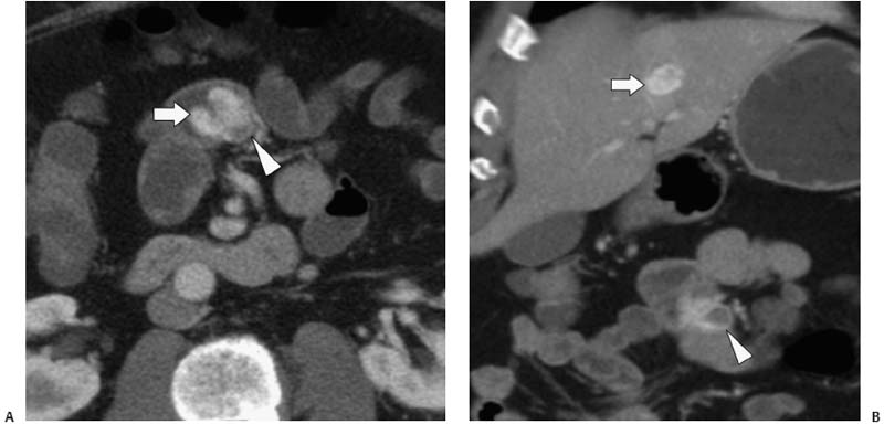
Case 18 A 70-year-old man presents to the gastroenterology clinic with weight loss, nausea, and vomiting. (A) Axial contrast-enhanced computed tomography (CT) shows an enhancing polypoid mass (arrow) extending into the lumen of the jejunum from a point of attachment to the bowel wall (arrowhead). (B) Coronal image shows an enhancing mass within the left lobe of the liver (arrow) in addition to the jejunal mass (arrowhead) with low-density central necrosis. • Gastrointestinal stromal tumor (GIST): This is the most likely diagnosis, indicated by a heterogeneous, lobulated polyp with regions of low attenuation after contrast administration. • Primary small-bowel adenocarcinoma:
 Clinical Presentation
 Imaging Findings
 Differential Diagnosis
![]()
Stay updated, free articles. Join our Telegram channel

Full access? Get Clinical Tree


