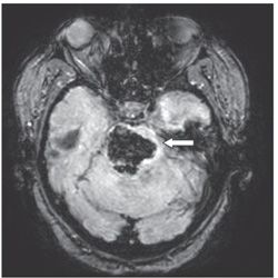
FINDINGS Figure 180-1. Axial NCCT through the pons. There is hyperdense material in the pons (arrow). There is mild expansion of the pons. Figure 180-2. Susceptibility MRI. There is corresponding hypointense signal characteristic of blood with expansion of the pons.
DIFFERENTIAL DIAGNOSIS Hypertensive hemorrhage, hemorrhagic tumor, hemorrhagic vascular lesion, amyloid angiopathy.
DIAGNOSIS Hypertensive brainstem hemorrhage.
DISCUSSION
Stay updated, free articles. Join our Telegram channel

Full access? Get Clinical Tree


