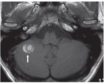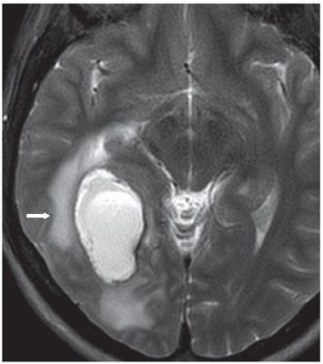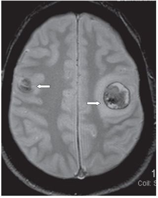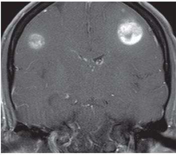


FINDINGS Figure 181-1. Axial NCCT through the corona radiata. There are four small well-circumscribed hyperdense masses (arrows) with surrounding hypodense edema in juxtacortical locations in the two hemispheres. The right parasagittal mass shows a hyperdense periphery surrounding a hypodense core. There were many other lesions with similar configuration elsewhere in the brain (not shown). Figure 181-2. Axial non-contrast T1WI through inferior posterior fossa. There is a right cerebellar peripherally placed “target” lesion with alternating rings of hyperintensity and hypointensity (arrow). There were many lesions with similar configuration elsewhere in the brain. Figure 181-3. Axial T2WI through the largest lesion in the right peritrigonal region. The mass is hyperintense with eccentric anterolateral target configuration. There is a hypointense thin smooth rim and surrounding hyperintense vasogenic edema. Other T2 lesions show a variegated intensity pattern. The trigone is compressed. Figure 181-4. Axial GRE through the centrum semiovale. Two lesions (arrows) with variegated mostly hypointense signal with hypointense rims and surrounding edema are present, one in each posterior frontal lobes in juxtacortical locations. Figure 181-5
Stay updated, free articles. Join our Telegram channel

Full access? Get Clinical Tree


