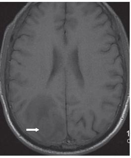
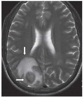
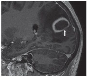
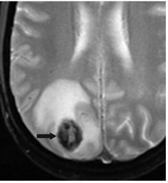
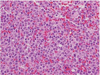
FINDINGS Figure 184-1. Axial DWI through the parietal lobes. There is a focus of hyperintensity (restricted diffusion) in the right parietal lobe (vertical arrow). Figure 184-2. Axial T2WI through mass. There is an ovoid right parietal isointense (with white matter [WM]) rim surrounding a core of mild hyperintensity (transverse arrow). There is surrounding hyperintense vasogenic edema (vertical arrow). Figure 184-3. Axial T1WI through the mass. The mass is hypo/isointense (arrow) with surrounding hypointense vasogenic edema. There is local mass effect. Figure 184-4. Sagittal post-contrast T1WI. There is a thick smooth ring enhancement of the lesion. The ring is attenuated inferoposteriorly (arrow). Edema extends into the occipital lobe inferiorly. Figure 184-5. Axial GRE through the mass. The mass shows a thick hypointense rim (arrow) surrounding a small hyperintense core with surrounding edema. Figure 184-6. Photomicrograph shows large lymphoid cells with atypical hyperchromatic nuclei; some with prominent nucleoli (monomorphic posttransplant lymphoproliferative disease [PTLD]/diffuse large B-cell lymphoma) (H&E stain).
DIFFERENTIAL DIAGNOSIS


Stay updated, free articles. Join our Telegram channel

Full access? Get Clinical Tree


