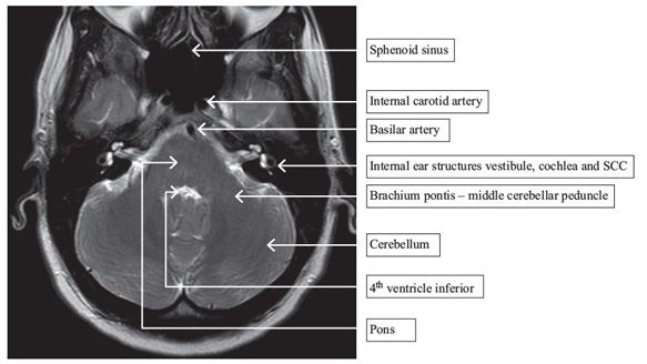
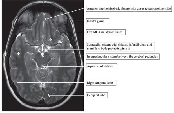
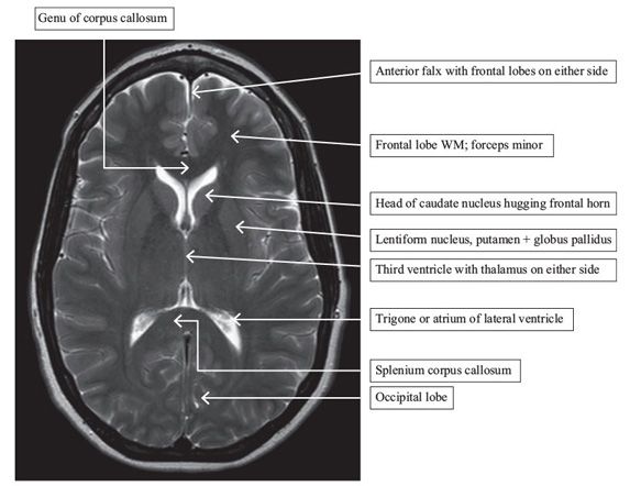
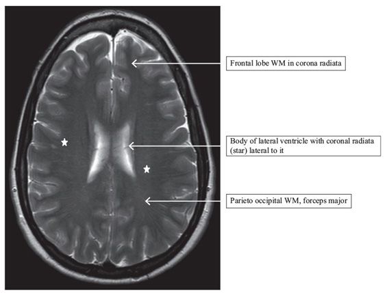
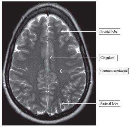
FINDINGS Images through different levels of the brain showing the structures as marked. Figure 185-1. Midline T1WI sagittal. Figure 185-2. Axial T2WI through the pons. Figure 185-3. Axial T2WI through the midbrtain. Figure 185-4


Stay updated, free articles. Join our Telegram channel

Full access? Get Clinical Tree


