Case 186
Case History
A 67-year-old woman is referred for new left breast lump. She was treated for left breast cancer 15 years ago with left lumpectomy, axillary node dissection, and radiation therapy.
Physical Examination
• left breast: diffusely erythematous, thickened skin; no dominant masses; slightly inverted nipple
• right breast: single enlarged axillary node; otherwise normal exam
Mammogram (Fig. 186–1)
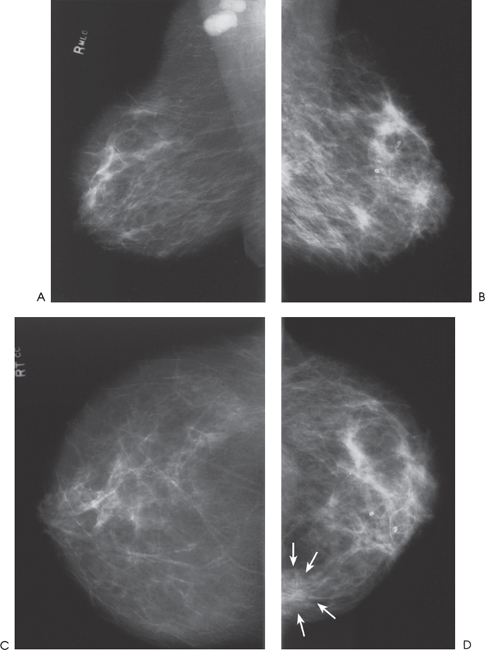
Figure 186–1. Old lumpectomy site is in the left axilla. There is diffuse increased density of the left breast with skin and trabecular thickening. An asymmetric density is present in the left medial breast (arrows). Multiple enlarged, dense nodes are present in the right axilla. (A). Right MLO mammogram. (B). Left MLO mammogram. (C). Right CC mammogram. (D). Left CC mammogram.
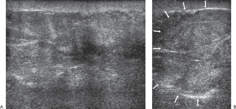
Ultrasound
Frequency
• 14 MHz (Figs. 186–2 and 186–3)