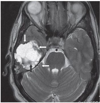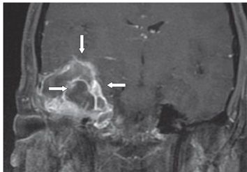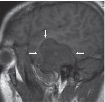


FINDINGS Figure 186-1. Axial cranial NCCT through the middle cranial fossa bone window setting. There is a 5 cm × 4.5 cm thin rim expansile mass in the right temporal fossa (star) arising from the petrous bone. It has irregular internal bony septations. The content is hypodense on brain window (not shown). Figure 186-2. Axial T2WI through the mass. The mass is homogeneously hyperintense except for the septations and the rim which are hypointense (arrows). The content follows cerebrospinal fluid (CSF) intensity on all sequences including DWI and ADC maps. There is no significant abnormality or edema of the surrounding brain. Effusion of the right mastoid air cells is present (not shown). Figure 186-3
Stay updated, free articles. Join our Telegram channel

Full access? Get Clinical Tree


