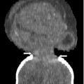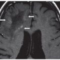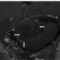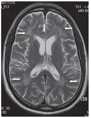
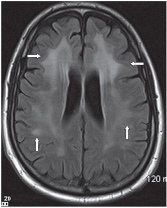
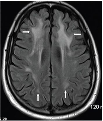
FINDINGS Figure 187-1. Axial FLAIR through the temporal lobes. There is bilateral anterior temporal lobes white matter (WM) hyperintensity (arrows). Figure 187-2. Axial T2WI through the basal ganglia. There is confluent bilateral symmetrical WM hyperintensity around the frontal and occipital horns extending from the ventricular walls to the subcortical regions (arrows). The frontal WM hyperintensity extend across the genu and anterior body of the corpus callosum (vertical arrow). This T2WI captures the degree of brain volume loss which is mild in this case. Figures 187-3 and 187-4. Axial FLAIR through the corona radiata and centrum semiovale, respectively. There is predominant symmetrical bilateral frontal lobes confluent WM hyperintensity extending from ventricular wall to subcortical regions (transverse arrows). The lesions are more subcortical, smudgy, and not as confluent in the parietal and occipital WM with relative sparing of the deep WM (vertical arrows). It is noted that there is no mass effect, and the post-contrast images (not shown) do not show areas of contrast enhancement.
DIFFERENTIAL DIAGNOSIS
Stay updated, free articles. Join our Telegram channel

Full access? Get Clinical Tree


