Case 188
Case History
A 35-year-old woman is admitted for severe disseminated encephalomyelitis of unknown origin. She is comatose.
Physical Examination
• unresponsive patient
• right breast: palpable lump in upper outer quadrant
• left breast: normal exam
Ultrasound
Frequency
• 10 MHz
Mass
• margin: ill defined
• echogenicity: hypoechoic
• retrotumoral acoustic appearance: bilateral edge shadowing
• shape: lobulated (Fig. 188–1)
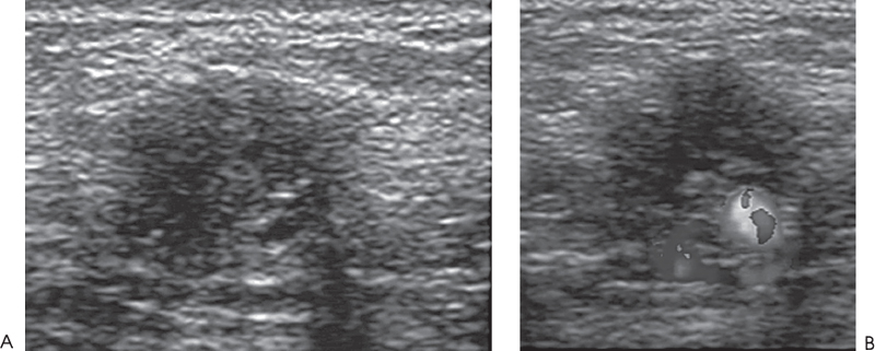
Figure 188–1. The palpable lump corresponds to a lobulated hypoechoic mass. (A). Right radial breast sonogram. (B). Right radial color Doppler breast sonogram (see Color Plate 188–1B).
Other Modalities (Fig. 188–2)


Stay updated, free articles. Join our Telegram channel

Full access? Get Clinical Tree


