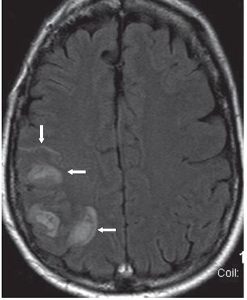
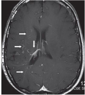
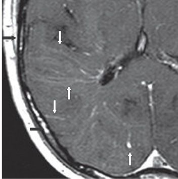
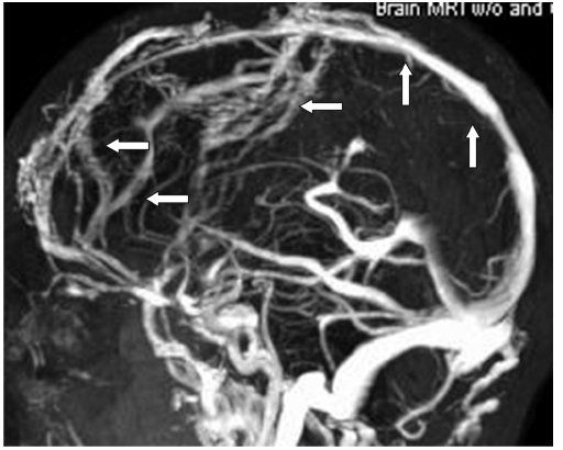
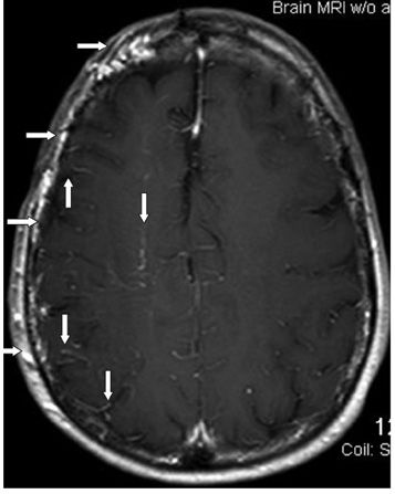
FINDINGS Figure 188-1. Sagittal contrast-enhanced MRV of the head. There is occlusion of major portion and irregularities of the superior sagittal sinus (SSS) consistent with SSS thrombosis (transverse arrows). There is extensive collateral venous drainage in the frontal region (vertical arrows). Figure 188-2. Axial FLAIR through the centrum semiovale. There are multifocal right frontoparietal subcortical hemorrhages (transverse arrows) with local mass effect. Right posterior frontal sulcal hyperintensity suggests subarachnoid hemorrhage (SAH) or leptomeningeal collateral (vertical arrow). Figure 188-3. Axial post-contrast T1WI through the lateral ventricles. There are multiple contrast-enhancing leptomeningeal vessels (transverse arrows) extending from the cortex through the white matter (WM) to the large right ependymal/thalamostriate vein (vertical arrow). Figure 188-4


Stay updated, free articles. Join our Telegram channel

Full access? Get Clinical Tree


