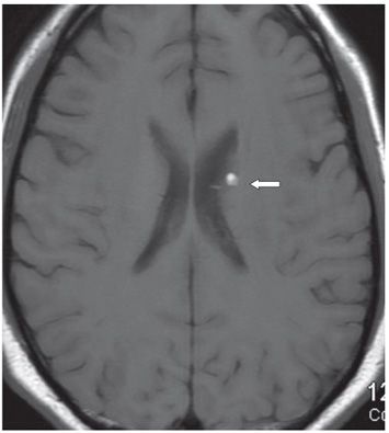
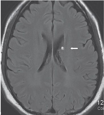
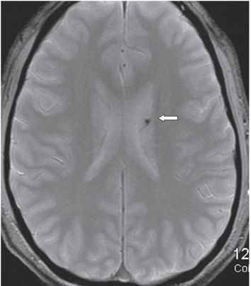
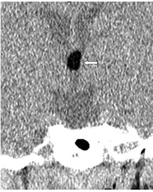
FINDINGS Figure 189-1. Axial NCCT through the superior lateral ventricles. There is a small fat hypodensity surrounded by calcification superiorly in the left lateral ventricle. The fat measures −62 HU compared with −1,018 HU in the air containing sphenoid sinus. Figures 189-2 and 189-3. Axial FLAIR and T1WI, respectively, on follow-up MRI demonstrate a small round hyperintensity in the location of the fat (arrow). Figure 189-4. Axial GRE through same level. There is a focal hypointensity in the location of the fat (arrow). Figure 189-5. Coronal NCCT through the third ventricle in another patient. There is an ovoid homogeneous fat hypodensity within the third ventricle (arrow) measuring −125 HU.
DIFFERENTIAL DIAGNOSIS


Stay updated, free articles. Join our Telegram channel

Full access? Get Clinical Tree


