 Clinical Presentation
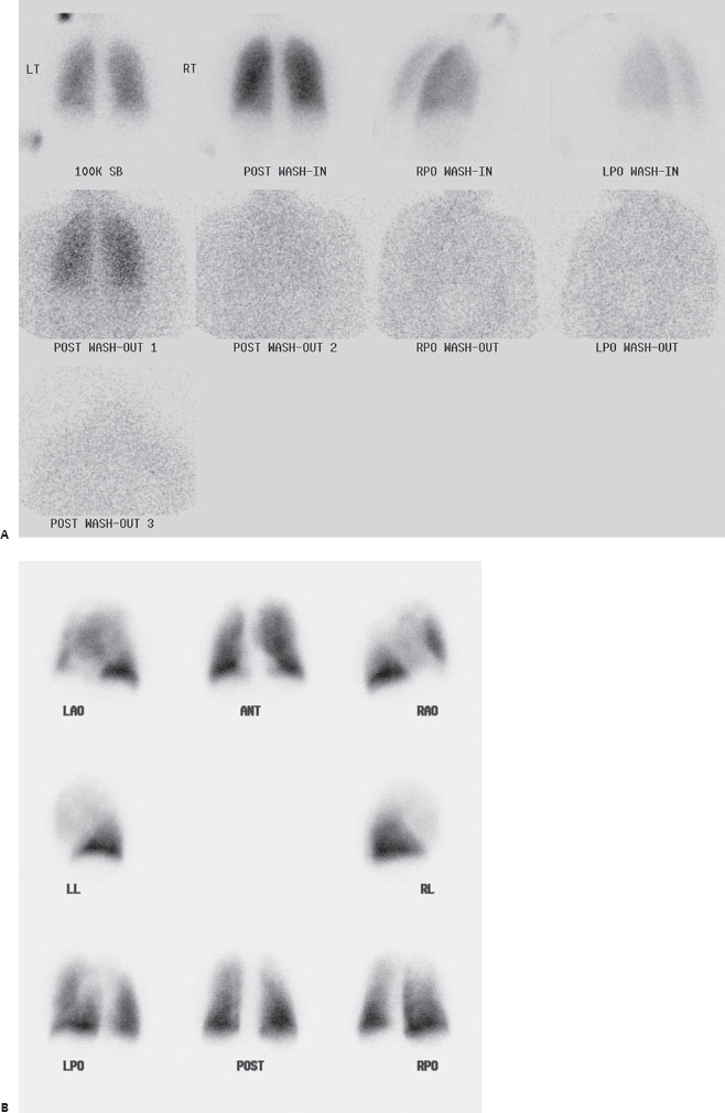
A 35-year-old woman with recent shortness of breath. Chest radiographs were unremarkable.

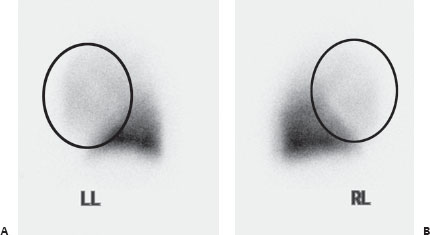
(A,B) Tc99m MAA perfusion images demonstrate significantly decreased perfusion to the upper lobes bilaterally, most dramatically on the lateral views (circles). Xenon 133 ventilation images are normal, so these are mismatched perfusion abnormalities.
 Differential Diagnosis
• Artifact of upright Tc99m-MAA injection: Uniform, symmetric, markedly decreased perfusion to the upper lobes with normal ventilation makes this the most likely diagnosis.
• High probability for pulmonary embolism (PE): This also appears as large, mismatched perfusion defects. However, the defects are rarely this symmetric and most commonly include the lower lobes as well (lower lobes receive the most blood flow).
• Asthma/chronic obstructive pulmonary disease: This can commonly demonstrate symmetric upper lobe perfusion abnormalities. However, these will be secondary to matched upper lobe ventilation abnormalities.


Stay updated, free articles. Join our Telegram channel

Full access? Get Clinical Tree


