Courtesy of M. Castillo MD.

Courtesy of M. Castillo MD.

Courtesy of M. Castillo MD.

Courtesy of M. Castillo MD.
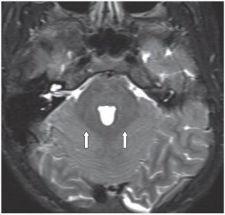
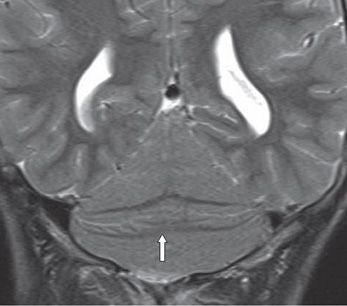
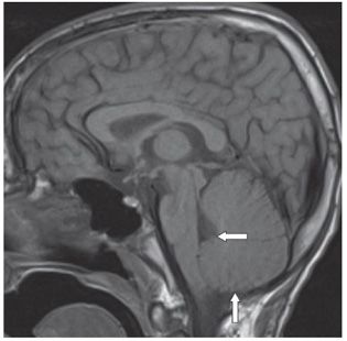
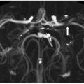
FINDINGS Figure 19-1. Axial T2WI through the lower cerebellum. There is a single cerebellum without a midline fissure. The folia are oriented transversely, and there is continuity of the cerebellar white matter (WM) across the midline (arrows). There is no distinct vermis. Figure 19-2. Axial T2WI through the fourth ventricle. There is fusion of bilateral middle cerebellar peduncles and dentate nuclei around the deformed fourth ventricle (arrows). There is no obvious nodulus. There is lack of midline fissure in the single cerebellar hemisphere. Figure 19-3. Coronal T2WI through the cerebellum. There is transverse orientation of the folia and WM (arrow). The cerebellum is pear shaped. Figure 19-4
Stay updated, free articles. Join our Telegram channel

Full access? Get Clinical Tree


