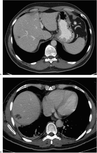
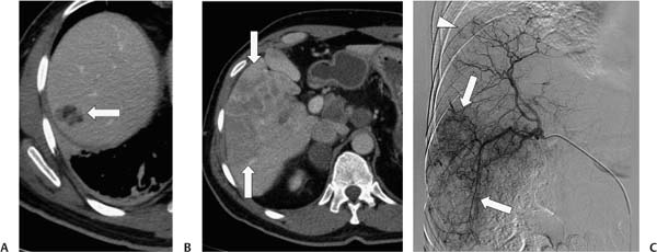
Case 19 A 54-year-old woman presents with nausea and weight loss. The second image was obtained 14 months after the first. (A) Contrast-enhanced computed tomography (CT) obtained after 14 months shows enlargement and cranial extension of the lesion (arrow) in the right lobe of the liver. The lesion is heterogeneous, with foci of fat attenuation as well as slightly hypoattenuating soft tissue. (B) More caudal slice shows a large, heterogeneous mass (arrows) on the 14-month follow-up study. (C) Selected angiogram shows extensive neovascularity in the right lobe (arrows), with a focus of neovascularity in the region of the lesion seen on CT (arrowhead). This study was obtained in preparation for chemoembolization. • Hepatocellular carcinoma (HCC): This is the principal diagnostic consideration for a growing mass in the liver with patchy macroscopic fat. The rest of the entities on this list are much less common but mentioned for discussion purposes.
 Clinical Presentation
 Imaging Findings
 Differential Diagnosis
Stay updated, free articles. Join our Telegram channel

Full access? Get Clinical Tree


