The Cerebellar Arteries
19.1 Case Description
19.1.1 Clinical Presentation
This 49-year-old man presented to medical attention with a large subarachnoid hemorrhage. CTA failed to demonstrate a source for the subarachnoid hemorrhage, and the patient was referred for diagnostic catheter angiography. This initially also failed to demonstrate a definitive source for the subarachnoid hemorrhage. Given the large quantity of subarachnoid blood, repeat vascular imaging was recommended in short course.
19.1.2 Radiologic Studies
See ▶ Fig. 19.1, ▶ Fig. 19.2.
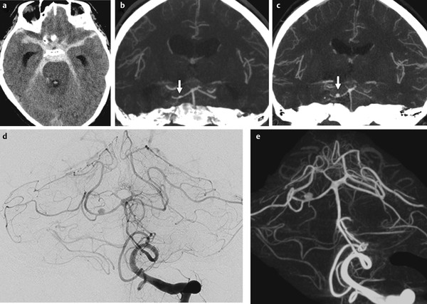
Fig. 19.1 Unenhanced axial CT (a) shows diffuse subarachnoid hemorrhage. Initial CTA in coronal view (b) was read as normal, although in retrospect and in light of the second CTA (c), a focal irregularity in the distal SCA could be appreciated that after 1 week had become more apparent and enlarged (arrows) to a fusiform outpouching. Left VA angiogram in anteroposterior (AP) view (d) and three-dimensional rotational reconstruction (e) demonstrate the fusiform aneurysm with adjacent proximal narrowing and which was therefore presumed to be of the dissecting type. Case continued in ▶ Fig. 19.2.
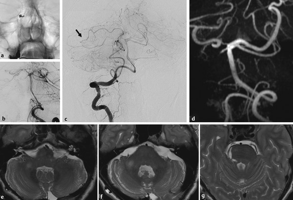
Fig. 19.2 Occlusion of the aneurysm and the parent vessel was performed (a,b), given the nature of the disease. On the control right VA angiography (c), there is collateralization from the vermian branch of the PICA to the medial vermian branch of the SCA (arrow). Follow-up time-of-flight MRA (d) demonstrated complete obliteration of the aneurysm. T2-weighted MRIs (e,f,g) demonstrate some small areas of ischemia in the brachium pontis, the superior cerebellar peduncle, and the dorsal tegmentum.
19.1.3 Diagnosis
Dissecting aneurysm of the distal medial division of the superior cerebellar artery (SCA).
19.2 Embryology and Anatomy
The cerebellum is essentially supplied by three pairs of arteries that are in balance with each other; namely, the SCA, the anterior inferior cerebellar artery (AICA), and the posterior inferior cerebellar artery (PICA). However, these arteries have distinctly different embryological origins: the SCA belongs to the anterior circulation, as it originates from the caudal ramus of the carotid artery, distal to the trigeminal artery; the AICA develops from the longitudinal neural system, a forerunner of the basilar artery; and the PICA, the latest acquisition to the cerebellar supply, can be regarded as a radiculopial artery that annexed a cerebellar territory when the cerebellum rolled over the myelencephalon.
The PICA is highly variable in both its origin (see Case ▶ 18) and territorial supply. Common variations include unilateral agenesis/hypoplasia, double or duplicated origin, and extracranial or epidural origin. The territorial supply of the PICA varies with its origin and branching arrangement. It can be absent in up to 26% of patients, and if so, its territory is typically annexed by a dominant AICA through the lateral medullary anastomotic network in the pontomedullary sulcus. An alternative supply of the PICA territory is the contralateral PICA, which can then lead to a bihemispheric supply. This is less common than an AICA–PICA complex, as the vascular territory has to be annexed by an artery that has to cross the midline. Intradural arteries can cross the midline via a commissure (e.g., bihemispheric supply by a single pericallosal artery) or a midline structure, such as the vermis, which permits the formation of a vessel that is not constrained to a single lateralized territory.
Stay updated, free articles. Join our Telegram channel

Full access? Get Clinical Tree


