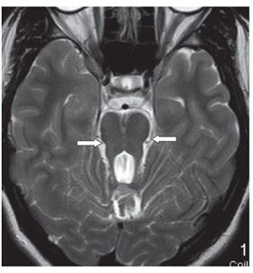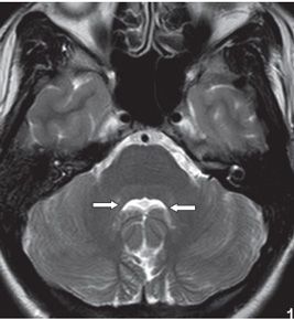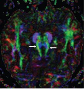


FINDINGS Figure 190-1. Sagittal MRI T1WI. There is a high-riding fourth ventricle (star) at the junction of the midbrain and the pons with obvious cerebellar vermis volume loss (arrow). The superior fourth ventricular velum is almost horizontal. Figure 190-2. Axial T2WI through the superior cerebellar peduncle demonstrates the molar tooth sign or malformation (MTS or MTM) (arrows). Figure 190-3. Axial T2WI inferior to the MTS. There is batwing appearance of the inferior fourth ventricle (arrows). Figure 190-4. DTI color directional map through MTS. There is thickening and horizontal disposition of the superior cerebellar peduncles (arrows).
DIFFERENTIAL DIAGNOSIS Joubert syndrome (JS), cerebellar vermian atrophy or hypoplasia, pontocerebellar atrophy.
DIAGNOSIS JS and Joubert syndrome-related disorder (JSRD).
DISCUSSION
Stay updated, free articles. Join our Telegram channel

Full access? Get Clinical Tree


