CASE 191
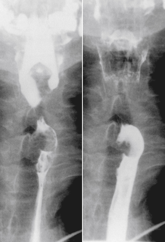
History: A 79-year-old man presents with dysphagia.
1. What should be included in the differential diagnosis of the imaging finding shown in the figure? (Choose all that apply.)
2. What is the most common benign neoplasm of the esophagus?
3. What is the most common clinical presentation of a patient with a fibrovascular polyp of the esophagus?
Stay updated, free articles. Join our Telegram channel

Full access? Get Clinical Tree


