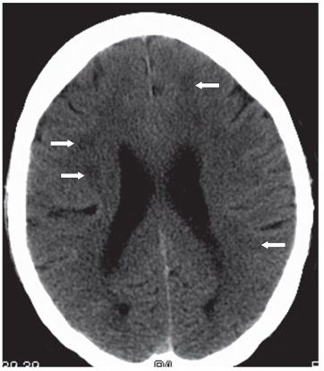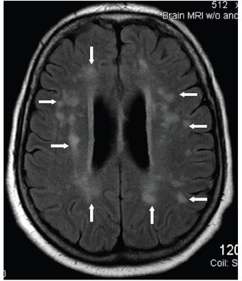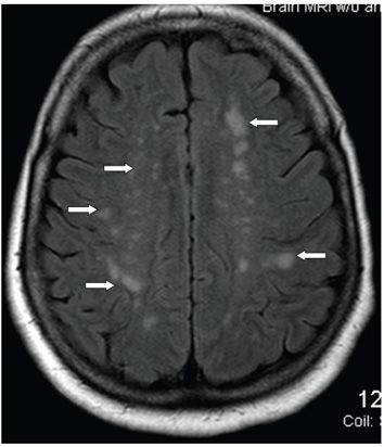


FINDINGS Figure 191-1. Axial NCCT through the basal ganglia. There is mild prominence of the lateral ventricles and sulci compatible with mild global brain volume loss. Multifocal bilateral almost symmetrical periventricular and subcortical white matter (WM) smudgy hypodensities (arrows) are present mainly in the frontal lobes. Figure 191-2. Axial NCCT through the lateral ventricles. There are multifocal poorly marginated hypodensities in bilateral deep and subcortical WM (arrows point to some of them). Figures 191-3 and 191-4. Axial FLAIR MRI through the corona radiata and centrum semiovale, respectively. There are multiple mostly punctate to small bilateral discreet WM hyperintensities mostly in the deep and subcortical regions (transverse arrows) with some smudgy hyperintensities around the occipital and frontal horns (vertical arrows).
DIFFERENTIAL DIAGNOSIS Demyelinating lesions, vasculitis, chronic small vessel ischemic changes, leukoaraiosis, and migraine.
DIAGNOSIS Multifocal WM chronic small vessel ischemic changes—leukoaraiosis.
DISCUSSION
Stay updated, free articles. Join our Telegram channel

Full access? Get Clinical Tree


