Case 192
Case History
A 49-year-old woman presents with a red spot on her right breast.
Physical Examination
• right breast: tender mass present at the 1:30 position; diffuse erythema of the upper inner quadrant
• left breast: normal exam
Mammogram (Fig. 192–1)
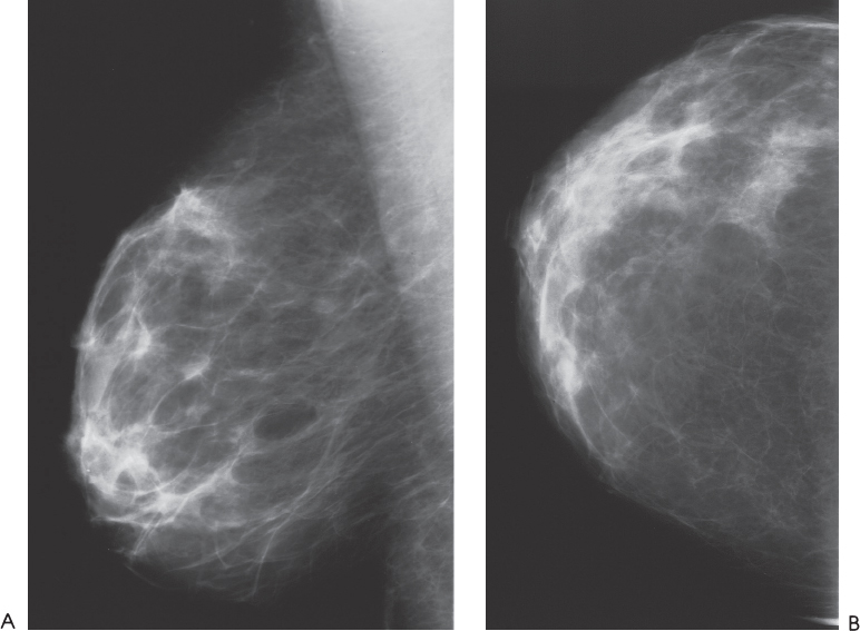
Figure 192–1. Right breast mammogram is normal. (A). Right MLO mammogram. (B). Right CC mammogram.
Ultrasound
Frequency
• 7 MHz
Mass
• margin: ill defined
• echogenicity: heterogeneous
• retrotumoral acoustic appearance: no shadowing
• shape: ellipsoid (Fig. 192–2)


Stay updated, free articles. Join our Telegram channel

Full access? Get Clinical Tree


