CASE 193
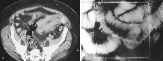
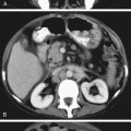
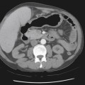
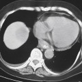
History: A 32-year-old woman presents with lower abdominal pain; a low hematocrit also is discovered.
1. What should be included in the differential diagnosis of the imaging finding shown in Figure A? (Choose all that apply.)
2. What is the most common cause of small bowel hemorrhage?
3. What finding on CT is most suggestive of small bowel hemorrhage?
A. Circumferential bowel wall thickening
Stay updated, free articles. Join our Telegram channel

Full access? Get Clinical Tree


