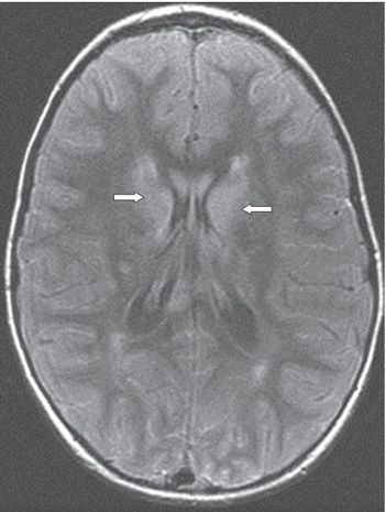
FINDINGS Figure 193-1. Axial FLAIR image through the thalami. There are asymmetric hyperintensities of both lentiform nuclei and thalami (arrows). Also note the high signal in posterior subarachnoid spaces. Figure 193-2. Axial FLAIR image cephalad to Figure 193-1. There is bilateral caudate nuclei hyperintensities (arrows). There is minimal white matter (WM) changes posterior to the occipital horns.
DIFFERENTIAL DIAGNOSIS Japanese encephalitis, West Nile virus encephalitis, Creutzfeldt-Jakob disease (CJD), bacterial meningitis, acute poliomyelitis, Guillain-Barré syndrome, and multiple sclerosis (MS).
DIAGNOSIS West Nile virus encephalitis.
DISCUSSION
Stay updated, free articles. Join our Telegram channel

Full access? Get Clinical Tree


