Case 194
Case History
A 42-year-old woman presents with new left breast lump.
Physical Examination
• left breast: ill-defined 5 cm area of thickening in the upper outer quadrant
• right breast: normal exam
Mammogram (Fig. 194–1)
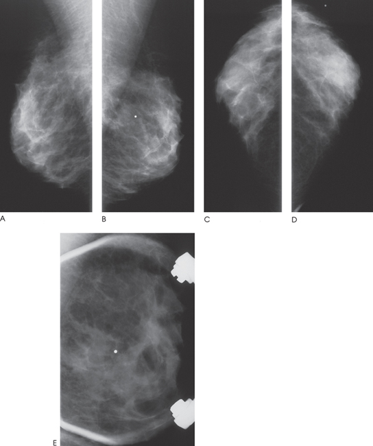
Figure 194–1. Bilateral mammograms demonstrate scattered fibroglandular densities. The palpable mass (labeled with a radiographic marker) is associated with an area of focal increased density in the left breast visible only on the MLO view (B,E). (A). Right MLO mammogram. (B). Left MLO mammogram. (C). Right CC mammogram. (D). Left CC mammogram. (E). Left MLO spot magnification mammogram of region of palpable mass.


Stay updated, free articles. Join our Telegram channel

Full access? Get Clinical Tree


