CASE 196
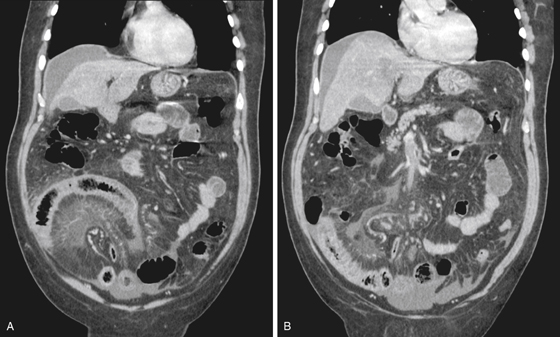
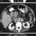
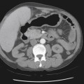
History: A 62-year-old man presents with a 4-day history of nausea and vomiting with a palpable mass in the right iliac fossa.
1. What should be included in the differential diagnosis of the imaging finding shown in Figure A? (Choose all that apply.)
2. What is the most common etiology for superior mesenteric venous occlusion?
3. What is the most specific imaging sign of superior mesenteric venous thrombosis?
Stay updated, free articles. Join our Telegram channel

Full access? Get Clinical Tree


