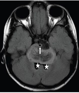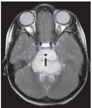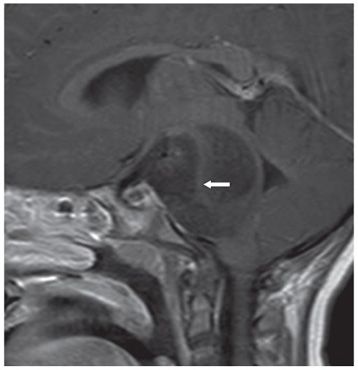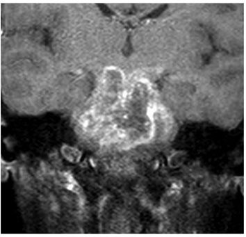


FINDINGS Figure 196-1. Axial NCCT through the pons. There is expanded hypodense pons obliterating all the surrounding cerebrospinal fluid (CSF) spaces including the fourth ventricle and wrapping around the basilar artery (arrow). Figure 196-2. Axial FLAIR through the pons. There is expansion of the pons with striated hypointense central portion and surrounding hyperintense periphery. There is a well-circumscribed hypointense component anteriorly and to the left of the basilar artery (arrow). There is effacement of the cerebellopontine angles (CPAs) and the prepontine cistern with compression of the fourth ventricle (stars). Figure 196-3. Axial T2WI through the pons. The mass is homogeneously hyperintense encasing the basilar artery (arrow). Figure 196-4. Post-contrast sagittal T1WI. There are two components to the non-contrast-enhancing hypointense mass, a posterior and an anterior component separated by an isointense S- shaped tissue (arrow). Figure 196-5
Stay updated, free articles. Join our Telegram channel

Full access? Get Clinical Tree


