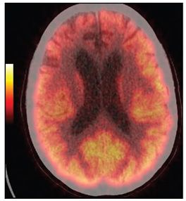
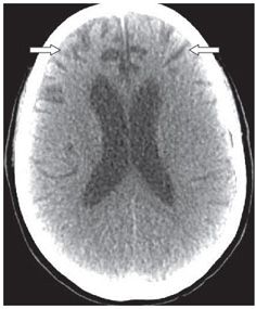
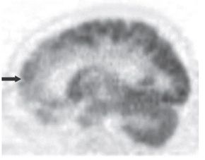
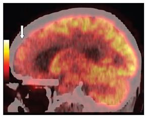
FINDINGS Figures 197-1. Top row. Axial F-18 FDG-PET images through the frontotemporal lobes. There is asymmetric mild to moderate decrease in FDG uptake in the frontal lobes (right greater than left) (arrows) Figure 197-2. The fused PET/CT and Figure 197-3 NCCT, show mild to moderate bilateral frontal volume loss (arrows). Figures 197-4 and 197-5


Stay updated, free articles. Join our Telegram channel

Full access? Get Clinical Tree


