CASE 198
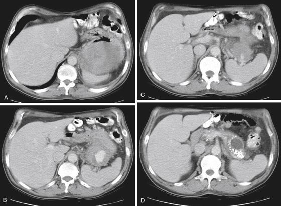
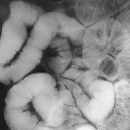
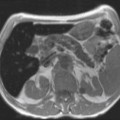
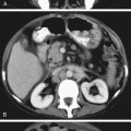
History: A 53-year-old man presents with hematemesis. Upper endoscopy identified an extramucosal mass distorting the gastric greater curvature.
1. What should be included in the differential diagnosis of the imaging finding shown in Figure A? (Choose all that apply.)
A. Colonic splenic flexure mass
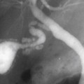
2. Which of the following statements regarding the differences between splenic artery aneurysms and pseudoaneurysms is true?
B. Pseudoaneurysms are usually clinically silent and diagnosed as incidental findings.
C. Most splenic artery aneurysms occur as a complication of pancreatitis.
D. Splenic artery pseudoaneurysms are more common than aneurysms.
3. What is the most common site of aneurysm formation in the abdomen?
Stay updated, free articles. Join our Telegram channel

Full access? Get Clinical Tree


