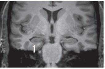
FINDINGS Figure 198-1. Coronal FLAIR through the hippocampus. There is asymmetry of the hippocampus. The right hippocampus is smaller than the left and hyperintense (arrow). Figure 198-2. Coronal T1WI through the hippocampus. Subtle decreased volume with flattened surface of the right hippocampus (arrow).
DIFFERENTIAL DIAGNOSIS Status epilepticus, mesial temporal sclerosis, choroidal fissure cyst, low-grade primary glioma, dysembryoplastic neuroepithelial tumor (DNET), posterior cerebral artery (PCA), or anterior choroideal artery (AChoA) infarct, hippocampal malrotation.
DIAGNOSIS Mesial temporal sclerosis (MTS).
DISCUSSION


Stay updated, free articles. Join our Telegram channel

Full access? Get Clinical Tree


